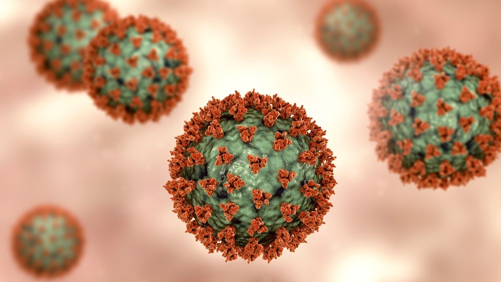
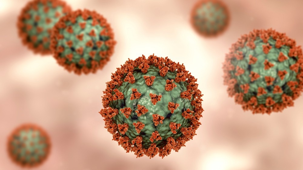
In a recent study posted to the bioRxiv* preprint server, researchers from Ireland used human bronchial epithelium models to evaluate the ability of a novel proprietary formulation called ViruSAL to inhibit severe acute respiratory syndrome coronavirus 2 (SARS-CoV-2) infections.

Background
SARS-CoV-2 infections begin in the epithelia of the upper airways but can progress into the lower airways. A large portion of severe coronavirus disease 2019 (COVID-19) morbidities and associated mortalities are due to interstitial pneumonia.
Ciliated bronchial epithelial cells in the upper airways express high levels of angiotensin-converting enzyme-2 (ACE-2) receptor and transmembrane serine protease 2 (TMPRSS2). SARS-CoV-2 spike proteins target the ACE-2 receptors to initiate viral entry, and cleavage of the spike protein by TMPRSS2 on the cell membrane further aids the process. Studies have shown that recent variants of concern, such as the Omicron variant, replicate faster in the ciliated epithelial cells than the earlier variants do.
Antivirals administered intranasally as prophylactics or to lower viral shedding have the potential to provide adjunct therapy along with vaccines and orally administered antivirals. Therefore, developing intranasally administered antivirals that inhibit SARS-CoV-2 infections in the ciliated bronchial epithelia could prevent infections from emergent variants.
About the study
The present study used VeroE6 cell lines and air-liquid interface (ALI) cultured human bronchial epithelial cells to assess the anti-viral efficacy of ViruSAL against SARS-CoV-2. They also used rat models to test whether ViruSAL interferes with the normal structure and function of mammalian cells in vitro and in vivo.
VeroE6 cells with SARS-CoV-2 infection were incubated in 0.5% to 3% ViruSAL to determine the concentration-dependent inhibition of SARS-CoV-2 by ViruSAL. Confocal imaging and transmission electron microscopy was used to examine the SARS-CoV-2 infected VeroE6 cells for viral presence and assembly.
Air-liquid interface cultured human bronchial epithelial cells were used to detect the inhibition of SARS-CoV-2 infection in the ciliated surface of the epithelial cells by different concentrations of ViruSAL. Additionally, the ability of ViruSAL to inhibit SARS-CoV-2 in pre- and post-infection ALI-cultured bronchial epithelial cells was also measured. Cell viability after ViruSAL treatment was assessed using 3-(4,5-dimethylthiazol-2-yl)-2,5-diphenyl-2H-tetrazolium bromide (MTT) assay.
The efficacy of ViruSAL against SARS-CoV-2 Alpha, Delta, and Omicron variants was measured using VeroE6 cells expressing TMPRSS2. Infectivity was quantified using 50% tissue culture infectious dose (TCID50) assays and quantitative reverse transcription polymerase chain reaction (qRT-PCR) to detect the SARS-CoV-2 nucleocapsid (N) gene. The safety and toxicity of ViruSAL were tested by intranasally administering ViruSAL on rat models.
Results
The results reported a concentration-dependent inhibition of SARS-CoV-2 by ViruSAL, with 3% ViruSAL resulting in undetectable levels of infection. Confocal imaging revealed a reduction in the expression of SARS-CoV-2 spike protein in infected VeroE6 cells treated with ViruSAL.
Transmission electron microscopy images showed no evidence of viral assembly in the endoplasmic reticulum Golgi intermediate compartments (ERGICs) of infected VeroE6 cells treated with 3% ViruSAL for two minutes while untreated and control-treated SARS-CoV-2 infected cells revealed virus-like particles in their ERGICs.
In ALI human bronchial epithelial cultures, ViruSAL displayed a time- and concentration-dependent reduction of infectivity, with 3% ViruSAL reducing SARS-CoV-2 infections to almost undetectable levels. ViruSAL also exhibited a concentration-dependent reduction of SARS-CoV-2 infections in post-infection ALI cultures of human bronchial epithelia.
ViruSAL was effective against all the tested variants of concern, with infections dropping to undetectable levels for SARS-CoV-2 Alpha and Omicron infections and very low levels of infectivity for Delta infection after treatment with 3% ViruSAL.
No adverse reactions or clinical signs of toxicity were reported in any rat models after the intranasal administration of ViruSAL. The body weights and appetites of all the animals were observed to be normal, and there was no difference in the lung weights of the euthanized test and control group animals.
Conclusions
Overall, the study revealed that ViruSAL effectively reduced SARS-CoV-2 infections in human bronchial epithelial cells cultured in ALI, in a concentration-dependent manner, with 3% ViruSAL completely inhibiting viral infection. ViruSAL was also effective in neutralizing infections in ALI-cultured cells post-infection.
Nasally administered ViruSAL displayed no evidence of toxicity in vitro, and no adverse reactions were seen in the rat models used to test the safety of ViruSAL in vivo. Furthermore, 3% ViruSAL effectively inhibited infections with SARS-CoV-2 Alpha, Delta, and Omicron variants, indicating its potential as a therapeutic agent against emergent variants.
*Important notice
bioRxiv publishes preliminary scientific reports that are not peer-reviewed and, therefore, should not be regarded as conclusive, guide clinical practice/health-related behavior, or treated as established information.
- Purves, K. et al. (2022) "A novel antiviral formulation inhibits SARS-CoV-2 infection of human bronchial epithelium". bioRxiv. doi: 10.1101/2022.10.05.510928. https://www.biorxiv.org/content/10.1101/2022.10.05.510928v1
Posted in: Medical Science News | Medical Research News | Disease/Infection News
Tags: AIDS, Angiotensin, Assay, Cell, Cell Membrane, Coronavirus, Coronavirus Disease COVID-19, covid-19, Efficacy, Electron, Electron Microscopy, Enzyme, Gene, Imaging, in vitro, in vivo, Mammalian Cells, Membrane, Microscopy, Omicron, Pneumonia, Polymerase, Polymerase Chain Reaction, Protein, Receptor, Respiratory, SARS, SARS-CoV-2, Serine, Severe Acute Respiratory, Severe Acute Respiratory Syndrome, Spike Protein, Syndrome, Tissue Culture, Transcription, Virus
.jpg)
Written by
Dr. Chinta Sidharthan
Chinta Sidharthan is a writer based in Bangalore, India. Her academic background is in evolutionary biology and genetics, and she has extensive experience in scientific research, teaching, science writing, and herpetology. Chinta holds a Ph.D. in evolutionary biology from the Indian Institute of Science and is passionate about science education, writing, animals, wildlife, and conservation. For her doctoral research, she explored the origins and diversification of blindsnakes in India, as a part of which she did extensive fieldwork in the jungles of southern India. She has received the Canadian Governor General’s bronze medal and Bangalore University gold medal for academic excellence and published her research in high-impact journals.
Source: Read Full Article
