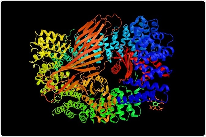
Mass spectrometry (MS) is considered to be a powerful method for quickly and efficiently identifying protein samples. Most crucial issues in the analysis of protein complex through MS include specificity of protein complex and contaminants in background protein.

Credit: ibreakstock/Shutterstock.com
Tandem affinity purification is the most recommended method to increase the protein complex specificity. Tandem affinity purification helps in improving the signal-to-noise ratio by purified sample generation. Stable isotopic protein labeling helps in identifying the background protein contaminants and binding partners.
Proteins usually combine with other components to form the complexes, stable or transient, which are involved in performing biological activities. In addition, some of the proteins may interact particularly with non-protein particles, such as RNA or DNA. These interactions are important for protein function. In this condition, MS method has been used to rapidly identify the complex component.
Procedure for analyzing protein using MS
Today, majority of the analysis of the protein sequence uses the MS method. Following are the some of the important steps involved in the protein analysis technique:
- The first step involved in protein analysis is purification, which is usually performed by affinity chromatography. After purification, the components of the protein complex can be separated using techniques that include isoelectric focusing, SDS-PAGE, or different types of separation method in 2D.
- Then the separated proteins are viewed by staining, and for further analysis, it can be recovered. By using trypsin, the separated proteins are digested proteolytically for creating the combination of the peptides that can be determined by mass spectrometer.
- There is no necessity for gel separation, if the sample has fewer complexities. Instead, MS can be directly applied to the digested protein complex. Finally, by using appropriate informatics tools, proteins in the sample are figured out by identified peptides recombination.
- Proteins are normally isolated on a reverse phase column using acetonitrile gradient. Generally, usage of trifluoroacetic in the solvent intercedes with ionization, which can be prevented by using acetic acid. Peptides that are ionized should be maintained in vapor state. This can be achieved by passing the column eluate that contains the solvent as well as peptides via a fine tip to create small droplets.
- The ionized peptides are maintained in vapor state, which occurs after solvent evaporation. The charged surfaces are allowed to transfer the ionized peptide into the MS. Liquid chromatography MS is a technique in which molecules are transferred into MS.
- Collisionally activated dissociation (CAD) or collision induced dissociation (CID) is an approach in which peptides are fragmented at peptide bonds by colliding them with gas molecules. From these peptides, calculate the mass of fragments. Since MS has two steps, such as mass of peptide fragment (MSn) and mass of peptide (MS/MS), sometimes there exist two or more steps for fragmentation.
- The process that contains these two steps is known as tandem MS. For determining the peptide arrangements, mass data of fragment can be utilized.
- Generally, data can be analyzed using computer comparing with identified sequences. Widely used program is SEQUEST. Sometimes data analysis is done manually for the newly introduced sequences and for the confirmation of some other main sequences.
Reason behind the incomplete protein sequence data
Predominantly, 70% of the protein sequence observed by viewing the corresponding peptide gives the successful analysis of the protein. Generally, 80% of the protein sequence coverage is sufficient to analyze the protein count involved in migration from a cell to another cell. Reasons behind the analysis which cannot identify all the amino acids are listed below:
- Improper digestion of protein.
- Because of the hydrophilic or small in nature peptide passes through the back stage column with the salt cannot be analyzed.
- Due to poor fragmentation, peptides are hydrophobic or too big, may not be removed from column, or sometimes too big for analyzing using a mass spectrometer.
- Ways of peptide fragmentation cannot be analyzed. Yet, several spectra are unexplained in the investigation.
Peptide Mass Fingerprinting (PMF)
This technique is used for the analysis of unidentified protein. Depending upon the database of the protein sequence, the complexity involved in identification of a protein is less. By using the Matrix Assisted Laser Desorption/Ionization instrument, mass of the peptides that are not fragmented, after the protein digestion with a specific proteinase or trypsin, can be estimated. Then the program has to compare the measurements that are calculated from the digested protein represented in a database with an obtained peptide mass.
This method is appropriate only for samples that contain a single protein, or a pair of proteins of the same abundance. If the proteins sequence is not available, then this method cannot be used. Finally, this method has been recommended as a route for analyzing samples from two dimensional gels.
Sources:
- https://www.ncbi.nlm.nih.gov/pmc/articles/PMC1665575/
- http://onlinelibrary.wiley.com/doi/10.1113/jphysiol.2004.080440/full
- med.virginia.edu/…/
- www2.chemistry.msu.edu/…/masspec1.htm
- http://www.geneontology.org/page/protein-complexes
- www.researchgate.net/…/288664230_Protein_complex_analysis_From_raw_protein_lists_to_protein_interaction_networks
- https://www.ncbi.nlm.nih.gov/pubmed/18370112
Further Reading
- All Mass Spectrometry Content
- What is Mass Spectrometry?
- Mass Spectrometry Applications?
- Coupling and Ionization in Miniature Mass Spectrometry
- Life Science Applications of the Top-Down Approach
Last Updated: Feb 26, 2019
Source: Read Full Article
