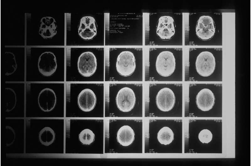
Objective assessment of irregularities in a tumor’s shape provides a means to evaluate its aggressiveness more effectively prior to an operation. This was the key finding made by ateam of physicians from Karl Landsteiner University of Health Sciences in Krems whose research focused on meningiomas, a form of tumor that affects the meningeal tissue in the brain. In a study published in the latest issue of the Journal of Neurosurgery, the research team demonstrated the high predictive value of the model they developed and named the surface factor. The model represents an objective and comparable parameter for the quantification of irregularities in tumor shape. Based on data from more than 125 patients, the study found a statistically significant correlation between a low surface factor (i.e. an irregular tumor surface) and a higher level of aggressiveness in the tumor.
Although meningiomas—tumors that develop in the meninges—are often benign, around 20% of cases are characterized by a higher degree of aggressiveness. Distinctions are drawn using the WHO classification, which grades tumors from I to III once they have been surgically removed and also determines the subsequent treatment regimen. However, pre-operative classification would be highly beneficial, as it would provide the surgeon with vital advance information on the best suited surgical strategy. A group of physicians from Karl Landsteiner University of Health Sciences in Krems (KL Krems) has now demonstrated that the factor they calculated provides precisely this type of information.
‘Superficial’ examination
“The starting point for our analysis is very simple,” explains Dr. Franz Marhold of the Clinical Department of Neurosurgery at KL Krems University Hospital in St Pölten. “Experience shows that tumors, especially meningiomas, with irregular surface tend to be more aggressive. This means we need a parameter for the objective quantification and comparison of irregularities in tumors. And this is exactly what we have achieved with the surface factor.”
Magnetic resonance imaging of a tumor provides the foundations for determining the surface factor (SF), which enables the surface area and volume of the tumor to be calculated using specialist software. In the second step, the surface area of a hypothetical sphere with the same volume as the tumor is calculated. Dr. Popadic, first author of the study, says, “Of all geometric forms, a sphere has the smallest surface area relative to its volume. In other words, it represents a sort of ‘idealised’ tumor with the smallest possible number of irregularities.” The SF is then calculated as the ratio of the sphere’s surface area to that recorded for the tumor. The more irregularities, the lowerthe SF.
Clear data set
In order to highlight the predictive value of the SF, Dr. Marhold, Dr. Popadic and their team collected data from 126 patients who had meningiomas removed at two neurosurgical centers in Austria between 2010 and 2018. The retrospective study demonstrated that distinctions can be made with a high degree of statistical accuracy between the WHO grades I-III using the SF. Further analysis showed that the SF is independent of other values and can therefore serve as an easily calculable pre-operative prognostic factor for the aggressiveness of a meningioma.
Source: Read Full Article
