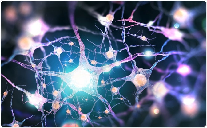
Regeneration of nerve cells involves either the repair or replacement of damaged nerve cells. While lower organisms possess an extensive capacity for neural regeneration, higher organisms, including humans, have limited ability to regenerate nerve cells.

Image Credit: Andrii Vodolazhskyi/Shutterstock.com
In humans, axons of the peripheral nervous system (PNS) are capable of regeneration, whereas those of the central nervous system (CNS) are currently viewed as incapable of regeneration.
This inability of the CNS to regenerate poses significant issues for the treatment of injury and disease of the nervous system.
Why do PNS axons regenerate, but CNS axons do not regenerate?
In higher animals like mammals, PNS axons regenerate after peripheral nerve damage spontaneously whereas, CNS axons do not regenerate after injury.
This difference in the regenerative capacity is attributed to the different types of glial cells present in these two systems: in the PNS, Schwann cells (SCs) promotes axonal regrowth whereas, in the CNS, regrowth is inhibited by oligodendrocytes and the formation of glial scars.
Glial scars inhibit nerve regeneration, significantly leading to a loss of function. Different molecules, such as transforming growth factors β-1 and β-2, interleukins, and cytokines, are released to promote glial scar formation.
Factors responsible for the regeneration of axons
Clearance of the debris
In the PNS, after peripheral nerve injury, the distal portion of the axon disintegrates from the soma within two days into small fragments by making small spheres of proteins called actin spheres, which breaks them down into smaller pieces.
These smaller fragments are then removed by Schwann cells and subsequently by macrophages. This process allows for the rapid clearing of axons and creates a favorable environment for axonal regrowth. In PNS axonal debris are cleared effectively within 2-3 weeks.
In CNS, after an injury, oligodendrocytes either die or remain unresponsive, they do not disintegrate the damaged axons in the central nervous system. They do not express vascular endothelial growth factor receptor 1(VEGFR1) like the Schwann cells.
However, genetic modification of oligodendrocytes can be used to induce VEGFR1 expression. A recent study showed that oligodendrocytes can be made to produce actin structures and disintegrate broken axon fragments like Schwann cells.
Upregulation of regeneration associated genes (RAGs)
An upregulation of regeneration associated genes (RAGs) in the PNS neurons has been observed following axotomy. Some of these RAGs are shown to play an important role in neurite outgrowth and regeneration.
These include c-Jun, activating transcription factor-3 (ATF-3), SRY-box containing gene 11 (Sox11), small proline-repeat protein 1A (SPRR1A), growth-associated protein-43 (GAP-43) and CAP-23.
CNS neurons cannot upregulate growth-associated genes; thus, even in the absence of inhibitors, they show limited regeneration capability.
The difference in the extracellular matrix
Another difference between the CNS and PNS is the basal lamina. Schwann cells secrete basal lamina composed of laminin, type IV collagen, and heparin sulfate proteoglycans (HSPGs), a substance required for myelination.
The abundance of basal lamina in the PNS and upregulation of pro-regenerative extracellular matrix molecules by Schwann cells promote PNS regeneration. Thus, the interaction of integrin-laminin activates the kinase enzymes, triggers intracellular signaling pathways, and promotes cytoskeleton rearrangement leading to axonal growth.
In contrast, oligodendrocytes secrete no basal lamina, and these molecules are mostly absent in the healthy CNS except at few places like pial surface.
However, some evidence suggests that astrocytes may upregulate growth-promoting extracellular matrix components such as fibronectin and laminin after injury. Still, these are overshadowed by the upregulation of chondroitin sulfate proteoglycans, which is inhibitory to the axon outgrowth.
Presence of inhibitors in the central nervous system
At the site of injury, reactive astrocytes are produced, and the up-regulation of inhibitory molecules takes place. These inhibitory molecules inhibit neurite outgrowth and contribute to the failure of neurodegeneration in the CNS.
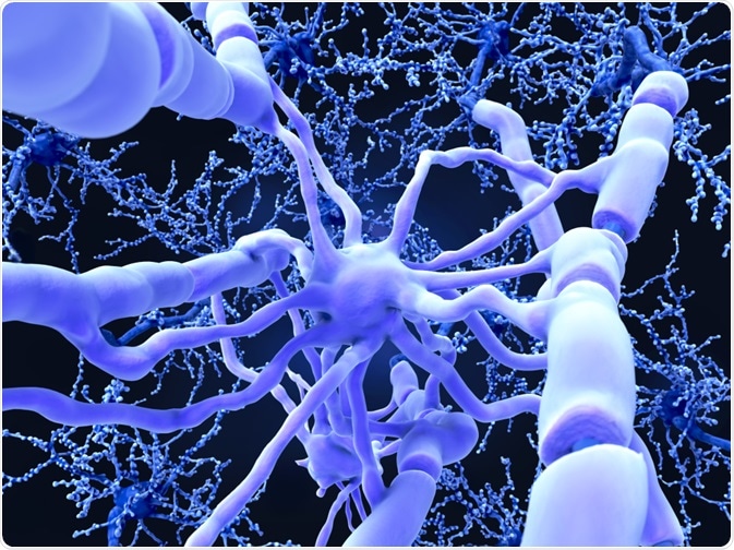
Axons in the nerves are sheathed in myelin, and damaged myelin forms the key to the regeneration of the axon. The myelin around the axon helps the nerve signals to pass quickly.

Image Credit: Juan Gaertner/Shutterstock.com
Myelin is essential for the function of the entire nervous system. Still, in case of injury, it hinders the repair process because of the presence of associated myelin integrins (MAI), a component of CNS myelin expressed by oligodendrocytes.
Using specific peptides or antibodies which block myelin inhibitory molecules and their receptors may improve the axonal regeneration and functional recovery.
Chondroitin Sulphate Proteoglycans
Chondroitin sulfate proteoglycans (CSPG) inhibit neuronal integrin (growth-promoting component) interactions with laminin. Neuronal Cell Adhesion Molecule (N-CAM) facilitates inhibitory effects of chemorepulsive Sema5a, limits the availability of calcium to neural molecules, and directly interacts with functional CSPG receptors on the neuronal surface.
Scientists have demonstrated that regeneration can be promoted either by enhancing growth promotion by heparin sulfate proteoglycans (HSPGs) or by using chondroitinase, which will digest CSPG.
Axonal mechanism
Axons communicate with their environment through cell surface adhesion, receptors, channels, and mechanosensitive molecules. Cell surface adhesion molecules enable growing axons to exert pressure on their environment and signal across the membrane. Besides, the growth factor receptor also drives axon signaling pathways.
Regenerating axons penetrate extracellular matrix, and integrin binds extracellular matrix glycoproteins to induce cell proliferation and axonal outgrowth leading to regeneration. Thus, the downregulation of integrins or its ligands leads to inhibition of axonal growth.
In the PNS, integrins required for axon growth are upregulated during regeneration, whereas in the CNS, integrin expression decreases with maturity or becomes selectively excluded from axons.
Thus in CNS, integrins are either in deficient levels or absent. Besides, inhibitory molecules such as NogoA and CSPGs present in CNS inactivates integrins leading to the nonavailability of appropriately activated integrins to enable axon regeneration.
To conclude, self-repair of the nerve is possible in the peripheral nervous system, but currently, no treatment for recovery of the central nervous system after an injury is discovered.
Attempts at nerve regrowth across the PNS-CNS transition have not been successful to date.
Sources
- Huebner, E. A., et al. (2009). Axon regeneration in the peripheral and central nervous systems. Results and problems in cell differentiation, 48, 339–351. https://doi.org/10.1007/400_2009_19
- Vaquié A1., et al. (2019). Injured Axons Instruct Schwann Cells to Build Constricting Actin Spheres to Accelerate Axonal Disintegration. Cell Rep.;27(11):3152-3166.e7. doi: 10.1016/j.celrep.2019.05.060.
Last Updated: Apr 15, 2020
Source: Read Full Article
