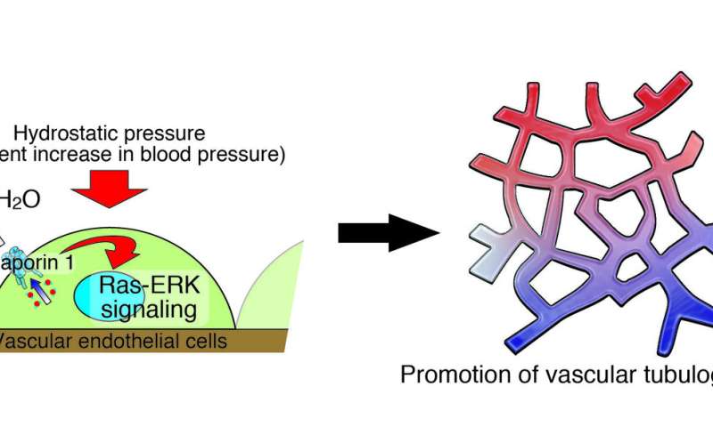
Blood vessels are the body’s transportation system, carrying oxygen and nutrients to cells and whisking away waste. But this vital mission can also be co-opted by tumors or break down as a result of disease, and this process has been poorly understood. An international collaborative team led by a scientist at Tokyo University of Agriculture and Technology (TUAT) in Japan has discovered a mechanism by which the growth of these vessels’ inner liner is stimulated, which may lead to therapeutics that could cut vessels off from tumors or help rebuild vessels in degenerative diseases.
The results were published on April 2 in Communications Biology, a Nature journal.
The researchers studied how the endothelial cells lining the inside of blood vessels respond to hydrostatic pressure, which is the pressure that forces blood through the vessels. The single layer of these cells forms a barrier between vessels and tissue, which controls fluid flow in the blood vessel walls.
“Blood vessels are constantly exposed to stimuli such as fluid shear stress, cyclic tensile force and hydrostatic pressure,” said Daisuke Yoshino, paper author and associate professor at the TUAT.
“Although the detailed mechanisms of cellular responses to shear stress and tensile force have been studied, we have not well understood how hydrostatic pressure affects vascular function.”
Yoshino and his team applied the pressure to endothelial cells, the equivalent of a person working out, to investigate how they sense hydrostatic pressure and convert it into biochemical signals, inducing vascular responses. They found that the increase in pressure triggered a water transfer through a channel called aquaporin 1, which activated a protein signaling channel called Ras-ERK. This process induced endothelial cell growth, indicating a clear connection between pressure stimulation and intracellular biochemical signals, according to Yoshino.
Source: Read Full Article
