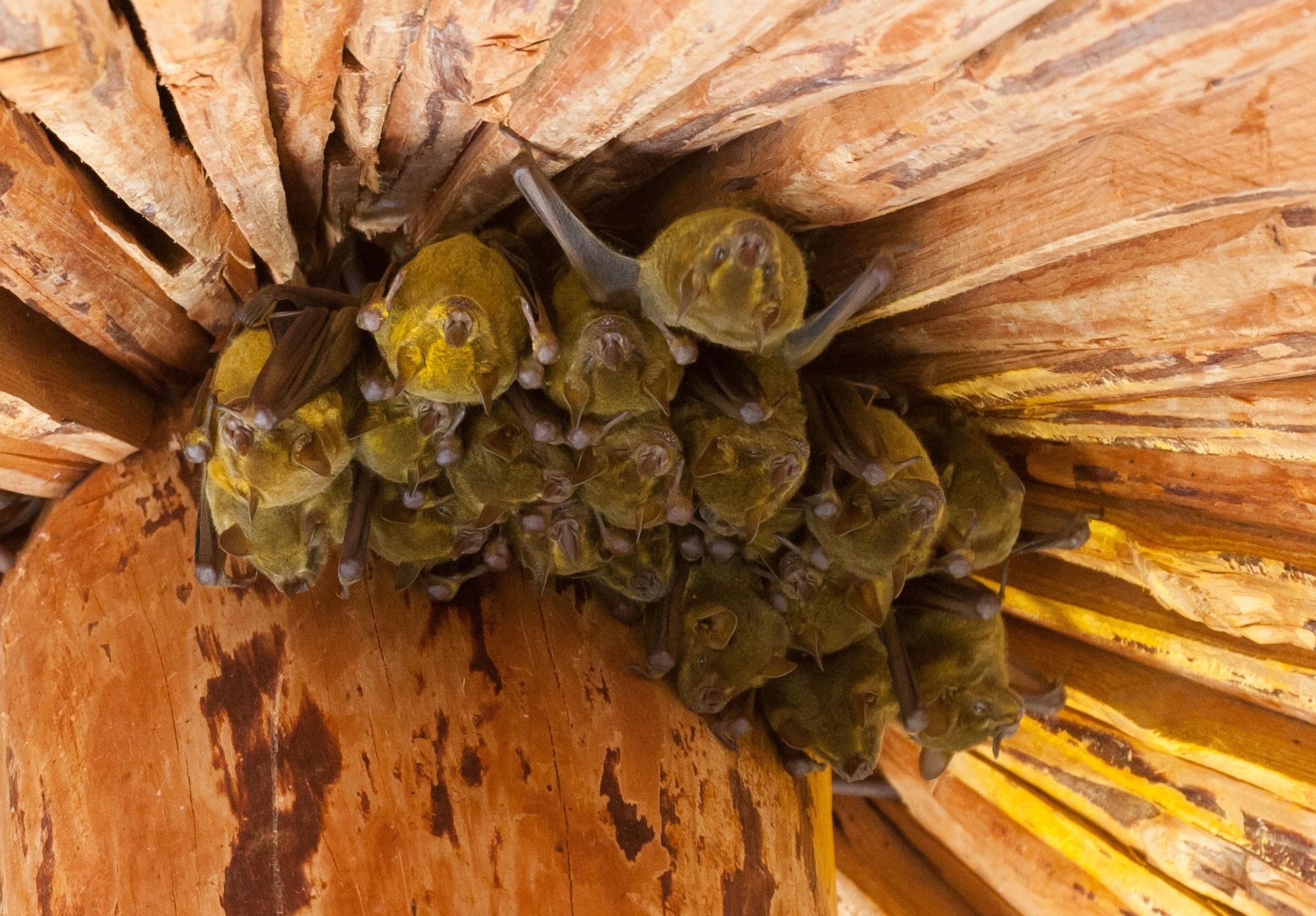
In a recent study posted to the bioRxiv* server, researchers examined the vulnerability of Jamaican fruit bats (Artibeus jamaicensis) to severe acute respiratory syndrome coronavirus 2 (SARS-CoV-2). They pursued evidence of how bats use their innate and adaptive immunity to engage SARS-CoV-2 or other coronaviruses (CoVs), although they do not have apparent clinical symptoms.
 Study: Regulatory T Cell-like Response to SARS-CoV-2 in Jamaican Fruit Bats Transduced with Human ACE2. Image Credit: Melinda Fawver / Shutterstock
Background
Natural (or wild) CoVs infect several bat species but have remained confined to their intestines, indicating fecal-oral viral transmission; however, studies have barely revealed the biology of SARS-related CoVs in bats. In all likelihood, Rhinolophus bat species harbored the ancestral SARS-CoV-2 that first spilled over into the bridge zoonotic host(s) and evolved further to transmit among humans giving rise to the coronavirus disease 2019 (COVID-19) pandemic.
It is unlikely that the ancestral SARS-CoV will ever be discovered because it has either become extinct, mutated or recombined with other sarbecoviruses. This also suggests that the initial Wuhan isolates of SARS-CoV-2 might behave differently in Jamaican fruit bats.
About the study
In the present study, researchers inoculated a replication-defective adenovirus (Adv) encoding human angiotensin-converting enzyme 2 (hACE2) or Adv/hACE2 in Jamaican fruit bats intranasally. Then they challenged them with SARS-CoV-2 after five days.
The researchers used a breeding colony of this bat species that they had established back in 2006 for performing infection studies with several viruses, including SARS and Middle Eastern Respiratory Syndrome (MERS)-CoVs. Initially, they maintained this bat colony in a free-flight facility. However, they shifted virus-infected bats to cages under biosafety level 3 (BSL-3) containment post-challenging with SARS-CoV-2 WA1 isolate. They sourced this strain from Biodefense and Emerging Infections (BEI) research resources repository and passaged it in Vero E6 cells to generate a stock.
Next, the team intranasally inoculated bats with 105 median tissue culture infectious doses (TCID50) of the virus. They held bats in a contralateral position to ensure delivery to the lungs and gastrointestinal tracts. Later, they collected their oral and rectal swabs on days two, four, seven, 10, 14, and 21 post-inoculation (PI) for viral ribonucleic acid (RNA) detection by quantitative polymerase chain reaction (qPCR).
Eventually, they euthanized bats for a necropsy and collected and froze portions of their lungs and intestine for RNA extraction and virus isolation. They used a SARS-CoV-2 envelope (E) gene detection kit for qPCR of RNA extracted from frozen bat tissues.
For in vitro studies, the researchers used coxsackievirus and adenovirus receptor (CAR) for transducing Jamaican fruit bat primary kidney epithelial (Ajk) cells with a replication-defective serotype Ad5 with a green fluorescent protein (GFP), at the multiplicity of infection (MOI) equals one for one hour. Since CAR shares 86% and 92% identity and similarity with its mouse counterpart, respectively, it was ideal for these experiments. The team monitored sustained fluorescence for several days. Later, they inoculated these cells with the virus without GFP for the SARS-CoV-2 challenge. For in vivo experiments, they inoculated five bats transduced with hACE2 intranasally using 2.5×10⁸ plaque-forming units (PFU) of SARS-CoV-2 WA1 isolate in 50 µl of Dulbecco's modified eagle medium (DMEM).
Twelve days later, they euthanized these animals to collect spleens for the activation-induced marker (AIM) test. This test uses a CD154 biomarker that identifies rare helper T (Th) cells reactive to antigen (fewer than 2% weeks after infection) by flow cytometry (FC). The rarity of these cells makes the assessment of ex vivo T cell responses challenging. Furthermore, the team also determined that a cluster of differentiation 4 (CD4)+ Th cells responded to SARS-CoV-2 infection in the hACE2-transduced bats.
Study findings
Omics eBook

The conserved ACE2 residues differ markedly among horseshoe bat species. For instance, Jamaican fruit bats have conserved 13 of 20 ACE2 residues (moderate). Due to low endogenous levels of ACE2 messenger RNA in the lungs of bats, the researchers could not detect SARS-CoV-2 in their lungs. Though the virus replicated in bats, it did so only for a few days, with bats showing no apparent clinical symptoms of disease or lost weight.
SARS-CoV-2 WA1 remained confined to the bat intestine, primarily, the Peyer's patches and the intestinal lamina propria's mononuclear cells. Also, they noted no apparent viral shedding in bat feces, and enzyme-linked immunosorbent assay (ELISA) sensitive to the SARS-CoV-2 nucleocapsid (NC) protein also failed to detect anti-SARS-CoV-2 antibody response. Overall, these results suggested that SARS-CoV-2 had poor susceptibility to bind Jamaican fruit bat ACE2 (AjACE2), making Jamaican fruit bat cells poorly permissible to the virus.
Perhaps, the Ajk cells lacked transmembrane serine protease 2 (TMPRSS2) that facilitates SARS-CoV-2 entrance through the plasma membrane. It is also possible that Adv transduction activated antiviral pathways in these cells that delimited SARS-CoV-2 replication. Another possibility is that SARS-CoV-2 infected cells via cathepsin L facilitate the endosomal evasion of the genomic RNA (gRNA). Nonetheless, transduction of hACE2 resulted in inefficient SARS-CoV-2 infection in other bat cell lines, none with a kidney origin. One of the cell lines produced infectious virions; however, those viruses could not complete exocytosis, likely because of tetherin, as observed using electron microscopy.
In hACE2-transduced bats challenged with SARS-CoV-2 WA1, the researchers detected low levels of viral RNA early in infection, likely due to limited Ad5/hACE2 penetrance in the distal ends of test animals from where the researchers extracted RNA. Histology results also showed clear pathology in hACE-transduced infected bat lungs, and the researchers readily detected antigens in their main stem bronchi. In SARS-CoV-2-challenged bats, low-titer antibody responses began on day 4. The titer of antibody responses remained low, even 21 days PI, likely due to a lack of IL-21 expression. Although, it is also possible that bats have robust innate immune responses that are less dependent on antibody responses.
The authors noted that the splenocyte cultures of three out of four bats stimulated for 24 hours with NC peptide library antigen showed no upsurges in interferon-γ (IFNγ) or interleukin-21 (IL-21) transcripts. In addition, there was a marked upsurge in CD154 expression on CD4+ Th cells (associated with severe COVID-19). Likewise, IL-10 and tumor necrosis factor-beta (TGFβ) were raised over two-fold, despite <2% of SARS-CoV-2 S-specific Th cells in the cell cultures.
Lastly, some subsets of regulatory T-cells (Treg) use these cytokines to suppress inflammatory responses. The authors noted a rise in the mRNA transcripts of Th cell markers CD4 and Cxcr4, suggesting that their activity might be less in bats or play a different role compared to humans. Thus, there were differences in Th cell behaviors in SARS-CoV-2-infected bats and humans.
Conclusions
With advancements in methods and reagents to study bat adaptive immunity, there will be more clarity regarding the roles that bat T cells play in immunity to viruses. Further studies should also focus on examining the role of individual T cells in bats using new techniques, such as FC, T cell culturing, or single-cell resolution RNA sequencing. In this context, as demonstrated by the current study, serotype 5 Adv vectors could prove beneficial. They could help dissect viral infections of bats, including from SARS-CoV-2 variants, and how their adaptive immune system responds to these viruses.
*Important notice
bioRxiv publishes preliminary scientific reports that are not peer-reviewed and, therefore, should not be regarded as conclusive, guide clinical practice/health-related behavior, or treated as established information.
- Regulatory T Cell-like Response to SARS-CoV-2 in Jamaican Fruit Bats Transduced with Human ACE2, Bradly Burke, Savannah Rocha, Shijun Zhan, Miles Eckley, Clara Reasoner, Amin Addetia, Juliette Lewis, Anna C Fagre, Phillida Charley, Juergen A Richt, Susan R Weiss, Ronald Tjalkens, David Veesler, Tawfik Aboellail, Tony Schountz, bioRxiv pre-print 2023; DOI: https://doi.org/10.1101/2023.02.13.528205, https://www.biorxiv.org/content/10.1101/2023.02.13.528205v1
Posted in: Medical Science News | Medical Research News | Disease/Infection News
Tags: ACE2, Adenovirus, Angiotensin, Angiotensin-Converting Enzyme 2, Antibody, Antigen, Assay, Biomarker, Bronchi, CD4, Cell, CLARITY, Coronavirus, Coronavirus Disease COVID-19, covid-19, Cytokines, Cytometry, Electron, Electron Microscopy, ELISA, Enzyme, Ex Vivo, Flow Cytometry, Fluorescence, Fluorescent Protein, Gene, Genomic, Histology, Immune System, immunity, in vitro, in vivo, Interferon, Interleukin, Interleukin-21, Kidney, Lungs, Membrane, Microscopy, Necrosis, Pandemic, Pathology, Polymerase, Polymerase Chain Reaction, Protein, Reagents, Receptor, Research, Respiratory, Ribonucleic Acid, RNA, RNA Extraction, RNA Sequencing, SARS-CoV-2, Serine, Severe Acute Respiratory, Severe Acute Respiratory Syndrome, Syndrome, Tissue Culture, Tumor, Tumor Necrosis Factor, Virus

Written by
Neha Mathur
Neha is a digital marketing professional based in Gurugram, India. She has a Master’s degree from the University of Rajasthan with a specialization in Biotechnology in 2008. She has experience in pre-clinical research as part of her research project in The Department of Toxicology at the prestigious Central Drug Research Institute (CDRI), Lucknow, India. She also holds a certification in C++ programming.
Source: Read Full Article
