
A new study published on the preprint server bioRxiv* proposes an integrative computational approach to model the structure of the severe acute respiratory syndrome coronavirus 2 (SARS-CoV-2) envelope.
Why study the structure of the viral envelope?
One of the different ways in which SARS-CoV-2 interacts with the host cell is through its envelope. To get a mechanistic understanding of this interaction, it is essential to have details on the structure of the viral envelope. Additionally, the surface of the viral envelope provides potential drug targets that can be used to design future therapeutic strategies.
The challenges in studying the structure of the viral envelope
SARS-CoV-2 belongs to a family of viruses called betacoronaviruses. These are enveloped positive-strand ribonucleic acid (RNA) viruses. Before the SARS-CoV-2 pandemic, there have been outbreaks of other pathogenic betacoronaviruses including SARS-CoV and the Middle East respiratory syndrome coronavirus (MERS-CoV).
Thus, several efforts have been made to elucidate the structural details of different viral proteins and their complexes. Even then, a detailed structure of the entire betacoronavirus particle remains unsolved.
SARS-CoV-2 particles are formed by four structural proteins including the spike (S) protein, which is required for receptor binding and membrane fusion, the membrane (M) protein, which is essential for membrane assembly, the E protein, which is present in low amounts, as well as the nucleocapsid (N) protein that protects the viral RNA. The N protein is responsible for the organization, packing, and protection of the viral RNA strand.
Comparatively, the S, M, and E proteins come together to form oligomers. The host cell membranes and these oligomers, together, form the viral envelope.
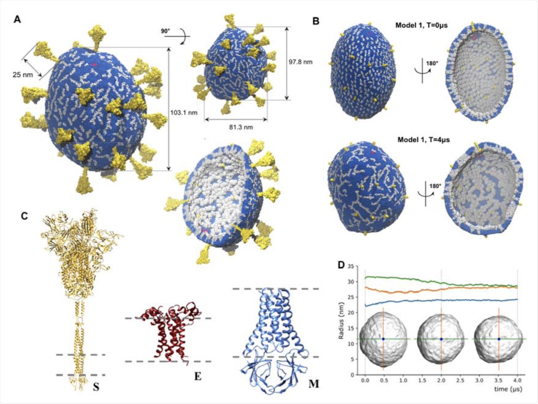 Structural characterization of SARS-CoV-2 viral envelope and its components: A. An envelope model obtained from configuration C1 (2 E pentamers, 25 S trimers, 1003 M dimers) and including full-length structures of S trimers after 1μs simulation run. Lipid molecules are depicted in sapphire blue, E pentamers in ruby red, M dimers in silver, and S trimers in gold. Principal diameters have values of 81.3 nm, 97.8 nm, and 103.1 nm. Height of the outer part of S protein is 25 nm. Surface of the envelope displays “filament” patterns formed by transmembrane domains of M dimers, while the internal part of the envelope shows tight packing of M dimers’ endodomains assemblies; B. Envelope model from configuration C1 using truncated S trimer structures at the start of the simulations (top) and after 4μs (bottom); C. Structural proteins S, E, and M representing the main structural building blocks of SARS-CoV-2 envelop in their physiological oligomeric states: S trimer, E pentamer, and M dimer. The grey dashed lines correspond to the membrane boundaries. The structures are shown in different scales. D. Change of the viral shape during the simulation defined through the principal gyration radii. The two largest principal radii converge to the value ∼28 nm while the third one converges to ∼24 nm. The actual diameters of the model after 4μs simulation were 103.1 nm, 97.8 nm, and 81.3 nm, respectively.
The viral envelope is flexible and lacks symmetry. The structures of E, M and S proteins have not been resolved experimentally. There are some structural insights available on S trimers and E pentamers, but there is no accurate model of the M dimer.
Moreover, the nature of the interactions between these three proteins is believed to be more complex than originally expected. The complexity and plasticity of the virus make it challenging to study the structure of the viral envelope.
Previous studies
Structural studies of several betacoronaviruses using electron microscopy and tomography have shown that the morphology of the viral envelope is conserved. However, there is a certain flexibility in its overall ellipsoidal shape.
It is estimated that the SARS-CoV-2 envelope contains 26±15 S trimers. While the number of M dimers in SARS-CoV-2 envelope is not known, they are estimated to be around 1,100 based on data from other betacoronaviruses.
Thus, M dimers are considered to be the most abundant protein in the envelope and its homodimers integrate into the virus' lipid bilayer. The E protein forms pentamers that are present in very few numbers on SARS-CoV-2 envelope.
These studies have provided information about the basic local and global geometric patterns that the structural proteins form on the envelope. However, this information is more general and low-resolution.
An integrative approach
In the current study, the scientists have taken an integrative approach to study the viral envelope of SARS-CoV-2. To this end, the researchers characterized S, M, and E proteins structurally, both in monomeric and homooligomeric forms. Additionally, the researchers determined the enrichment of specific lipids because of protein interactions using fingerprinting analysis.
Data was also collected from several sources to assemble the initial structural configuration of the envelope. Here, the researchers of the current study took data on the refined structures of the three homo-oligomers, their stoichiometry, the composition of the lipid bilayer, as well as the geometry and size of the envelope
Molecular dynamics simulations of several configurations were also conducted. Additionally, a structural and network analysis was also completed to unveil characteristic patterns of the structural proteins in the envelope assembly.
Taken together, this approach generated detailed models of the SARS-CoV-2 envelope by building on data available from previous studies.
Study results
According to this study, the SARS-CoV-2 M proteins are tightly and stably packed to form dimers. The monomers are also complementary in shape, which allows the natural tiling of multiple dimers into ‘filament’ structures. Thus, the M dimers agglomerate into large, filament-like, macromolecular assemblies that exhibit distinct molecular patterns.
The results of this computational study corroborate with previously published experimental data. Moreover, the structural and functional insights provided by this approach are beyond the purview of data obtained by a single experimental method.
Implications
This study provides a foundation for furthering our knowledge of the underlying molecular architecture of the entire SARS-CoV-2, the interplay between other protein oligomers, the role of orientation in virus-host interactions, and the molecular mechanisms of virus assembly. Furthermore, it can help in understanding the mechanistic determinants of the stability of the virus particle.
This automated computational protocol can be employed to study the envelope structure of other coronaviruses. This structural knowledge can also help in designing new antiviral compounds, as well as viral-like nanoparticles for novel vaccines, that can prevent the formation of the viral envelope.
*Important notice
bioRxiv publishes preliminary scientific reports that are not peer-reviewed and, therefore, should not be regarded as conclusive, guide clinical practice/health-related behavior, or treated as established information.
- Pezeshkian, W., Grünewald, F., Narykov, O., et al. (2021). Molecular architecture of SARS-CoV-2 envelope by integrative modeling. bioRxiv. doi:10.1101/2021.09.15.459697. https://www.biorxiv.org/content/10.1101/2021.09.15.459697v1.
Posted in: Device / Technology News | Medical Science News | Medical Research News | Disease/Infection News
Tags: Cell, Coronavirus, Coronavirus Disease COVID-19, Electron, Electron Microscopy, Lipids, Membrane, MERS-CoV, Microscopy, Morphology, Nanoparticles, Pandemic, Protein, Receptor, Respiratory, Ribonucleic Acid, RNA, SARS, SARS-CoV-2, Severe Acute Respiratory, Severe Acute Respiratory Syndrome, Syndrome, Tomography, Virus

Written by
Dr. Shital Sarah Ahaley
Dr. Shital Sarah Ahaley is a medical writer. She completed her Bachelor's and Master's degree in Microbiology at the University of Pune. She then completed her Ph.D. at the Indian Institute of Science, Bengaluru where she studied muscle development and muscle diseases. After her Ph.D., she worked at the Indian Institute of Science, Education, and Research, Pune as a post-doctoral fellow. She then acquired and executed an independent grant from the DBT-Wellcome Trust India Alliance as an Early Career Fellow. Her work focused on RNA binding proteins and Hedgehog signaling.
Source: Read Full Article