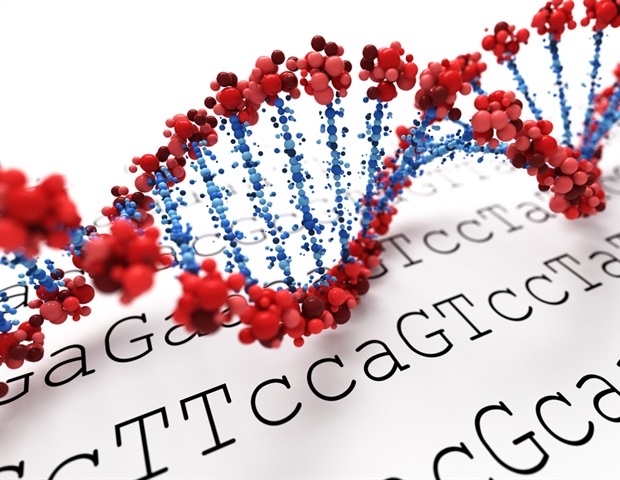
Optical genome mapping involves the extraction of very long DNA molecules, for example routinely collected blood samples or bone marrow material from patients. These long DNA molecules are labeled with dye molecules at more than half a million different positions in the entire human genome and are then moving through ultrathin nanochannels on a special chip. As the DNA molecules move through the nanochannels, a laser is used to make them visible and they are photographed using a fluorescence microscope. The images of the entire genome are then analyzed using bioinformatic analyses. "The aim is to identify and interpret changes in genetic regions that are relevant for the development of cancer," explains Dr. Wanda Gerding from the Bochum Department of Human Genetics.
Optical genome mapping thus facilitates genome-wide analysis of regions that are important for the classification and therapy of leukemias using one methodology. Furthermore, it also allows the identification of new relevant genomic regions and new genes.
Reliable and additional results
In the current study, the team compared the methodology to current standard diagnostics in patients with acute myeloid leukemia as well as myelodysplastic syndromes. The researchers showed that the results obtained by optical genome mapping methodology were concordant in 93 per cent of samples compared toa conventional methodology, the so-called cytogenetic karyogram, where whole chromosomes are visualized. In 67 per cent of the samples, it was even possible to obtain additional genetic information.
The methodology can thus not only detect structural changes in the genome more accurately, but also has the potential to become an important component of routine diagnostics for patients with leukemia.
As a further benefit, genome research can provide data and new insights for further research work in the field of tumor biology."
Dr. Wanda Gerding from the Bochum Department of Human Genetics
Cooperation partners
For the project, the Human Genetics Department at RUB, headed by Professor Huu Phuc Nguyen, cooperated with the Haematology, Oncology, Stem Cell and Immunotherapy Department of the Knappschaftskrankenhaus Bochum, headed by Professor. Roland Schroers, a member of the Centre for Haematooncological Diseases (ZHOE) at RUB, and Professor Peter Reimer from the Haematology, Internal Oncology and Stem Cell Transplantation Department at Evangelische Kliniken Essen-Mitte. The close scientific cooperation of both departments was ensured by Dr. Deepak Vangala, Dr. Wanda Gerding, Dr. Verena Nilius-Eliliwi (funded by the "Female Clinical Scientist" programme of the RUB Medical Faculty) and medical student Marco Tembrink (Human Genetics, medical doctoral scholarship holder from FoRUM, Medical Faculty of the RUB (FoRUM RUB). The project received a positive vote from the Ethics Committee of the RUB Medical Faculty (No. 20-7063).
Ruhr-University-Bochum
Gerding, W.M., et al. (2022) Optical genome mapping reveals additional prognostic information compared to conventional cytogenetics in AML/MDS patients. Cell Reports. doi.org/10.1002/ijc.33942.
Posted in: Genomics | Fluorescence
Tags: Blood, Bone, Bone Marrow, Cancer, Cell, CHIP, Diagnostics, DNA, Fluorescence, Genes, Genetic, Genetic Information, Genetics, Genome, Genomic, Haematology, Immunotherapy, Medicine, Microscope, Myelodysplastic Syndromes, Oncology, Research
Source: Read Full Article