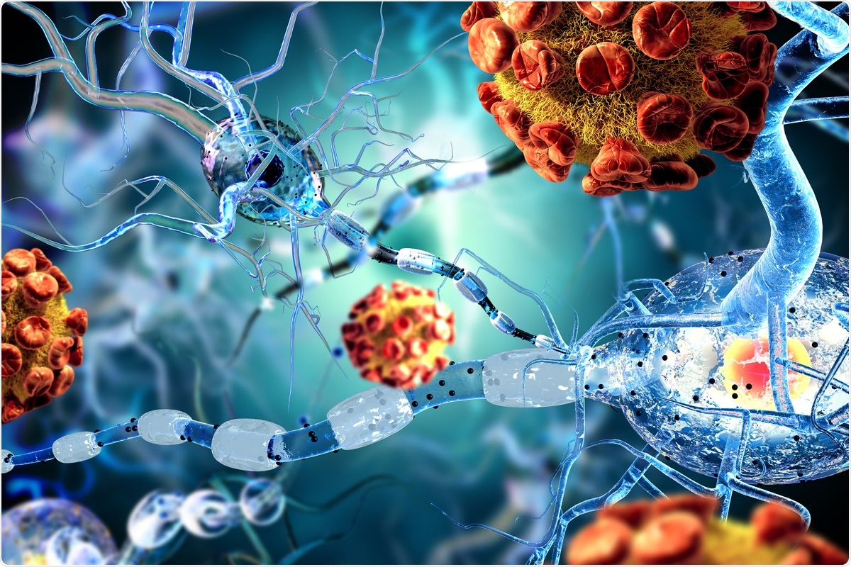
The coronavirus disease 2019 (COVID-19) pandemic has been associated with both short- and long-term neurologic complications, including stroke, brain fog and persistent tiredness.
A new study concludes that the effects of the severe acute respiratory syndrome coronavirus 2 (SARS-CoV-2) on the central nervous system are due to the endothelial injury and inflammation that this produces in the brain.

A preprint version of the study is available on the medRxiv* server, while the article undergoes peer review.
Study aims
Since the beginning of the pandemic, it has become clear that men are often more affected by COVID-19, with a higher likelihood of severe illness and a greater chance of death.
The current study focused on assessing brain injury markers (BIM) within 48 hours of hospitalization and at three months later.
BIMs are recognized as being valid indicators of injury to nerve cells and astrocytes, in human immunodeficiency virus (HIV) infection, sepsis and cardiac arrest. The current study focused on six, namely, glial fibrillary acidic protein (GFAP), neuron-specific enolase (NSE), S100B, ubiquitin carboxyl-terminal hydrolase isozyme L1 (UCHL1), Syndecan-1 and microtubule-associated protein 2 (MAP 2).
The scientists also examined levels of two markers of endothelial injury (Intercellular Adhesion Molecule 1, ICAM-1 and Vascular Cell Adhesion Molecule 1, VCAM-1) and of inflammation, in the form of cytokines or chemokines.
These were measured in hospitalized patients and in controls in a single hospital in Houston, Texas, USA. None of them had chronic lung, heart, neurological or psychiatric disease, cancer, or any disabling condition.
Increased markers of endothelial and brain injury
The researchers found that within 48 hours of hospitalization, that is, during the acute phase, patients had higher markers of brain injury like MAP2 and NSE than controls. The mean levels showed an increase of 60% to 145%, depending on the individual marker, relative to the controls.
Of these markers, MAP2 is a sign of dendritic injury, and was high at both acute and chronic time points. It has previously been shown to be high after traumatic brain injury and predicts long-term outcomes.
NSE is found in nerve cells and indicates damage. S100B is found in astrocytes and is high in traumatic brain injury and in strokes. Thus, this combination of BIMs shows combined nerve cell and astrocytic injury in COVID-19, worse in men than in women.
However, all markers had returned to normal at three months from hospitalization.
Markers of endothelial injury were also higher with acute infection, with the mean levels being two and three times higher than in controls, for ICAM1 and VCAM1, respectively. These were not assessed at three months.
The endothelial marker ICAM1 is released in response to IL-1b and TNFα. The effects are increased leukocyte adhesion, which reduces the barrier's integrity and promotes leakage from the blood vessels.
Cytokines and chemokines were also much higher, in some cases, in acute infection, but others showed a decrease. Of 38 chemokines and cytokines evaluated, seven were high, while two were low. Again, these reverted to normal levels at three months.
TNFα is a potent inflammatory mediator. Its elevation in this context indicates that the vascular injury is probably inflammatory in origin and not due to viral injury.
Brain injury due to inflammation and endothelial injury
The researchers carried out bioinformatics analysis to bring out possible associations between the markers. This showed that BIMs were strongly positive in their association with elevated or reduced cytokines/chemokines.
The pro-inflammatory cytokine interleukin-6 (IL-6) tended to be higher in men with COVID-19. People with higher IL-6 levels also had higher titers of other cytokines and BIMs.
Several other cytokines and chemokines, including IL10 and tumor necrosis factor-alpha (TNFα) were associated with brain injury as well. Thus, it appears that these signaling molecules drive inflammatory brain injury.
In other words, brain injury in acute COVID-19 is induced by systemic inflammation.
When analyzed by severity of disease, the BIMs showed no distinctions, but severe COVID-19 patients showed higher levels of sICAM1, an endothelial injury marker, and a couple of cytokines (SCD40L and IL1RA).
Greater inflammation linked to worse outcomes
The researchers found that the chemokine MIP1b (but no BIM or EIM) indicated a greater probability of adverse outcomes, that is, the functional status of the patient at discharge.
Raised markers in males
Among the BIMs, NSE was the only one that was raised in men relative to women. Four cytokines/chemokines were also elevated – IL10, IL15, IL8 and MIP1a, while four were near borderline significance.
Overall, men were more likely to show strong relationships between the levels of various BIM, EIM and cytokines. The pattern of correlation was different in men relative to women.
For instance, cytokines were correlated to BIMs in greater depth in men, indicating that inflammatory brain injury was more pronounced in this sex. Endothelial injury also appeared to be more pronounced in men, and showed a greater correlation with cytokines and BIMs.
What are the implications?
The findings showed that acute COVID-19 is linked to markers of brain injury, and these return to normal by three months. This is accompanied by signs of endothelial injury, which show strong positive associations with EIMs and several inflammatory cytokines.
However, men appeared to suffer worse brain injuries than women in acute infection, indicating that they are biologically susceptible to COVID-19-induced inflammation, endothelial damage, and brain injury.
The elevated BIMs suggest that there is CNS involvement in acute COVID-19 and the clinical ramifications need to be investigated urgently."
Earlier research showed that the blood-brain barrier is injured in COVID-19, and the viral spike protein may enter the CNS by this route. This injury is due to endothelial damage. Such damage has been documented by a study from the National Institutes of Health (NIH).
This damage may have been caused by direct viral injury, or because of the inflammation, or perhaps both. Further research will be required to understand the mechanism of such damage.
There were no obvious differences between the levels of these markers across grades of severity. This could be because acute-phase samples were all drawn from the same point of time (within 48 hours of admission). At this point, significant differences are unlikely to have emerged.
While long COVID has been extensively reported, the current study failed to show any elevations in BIMs, EIMs or cytokines, except in the case of MIP1a, at three months following hospitalization. This indicates that inflammatory injury is not ongoing, even though it may leave long-term sequelae on the survivors.
Men seem to develop a stronger inflammatory response to COVID-19, and to have more severe endothelial injury as a result. This fact must be kept in mind while treating male COVID-19 patients, even though the exact reasons for this difference from women with the same illness are as yet unclear.
The researchers conclude:
Further studies are required to clarify the exact mechanisms how COVID-19 induces inflammation and brain injury, sex-specific differences and whether these early increase in injury markers are associated with long-term neurologic outcomes."
*Important Notice
medRxiv publishes preliminary scientific reports that are not peer-reviewed and, therefore, should not be regarded as conclusive, guide clinical practice/health-related behavior, or treated as established information.
- Savarraj, J. et al. (2021). Markers of brain and endothelial Injury and inflammation are acutely and sex specifically regulated in SARS-CoV-2 infection. medRxiv preprint. doi: https://doi.org/10.1101/2021.05.25.21257353, https://www.medrxiv.org/content/10.1101/2021.05.25.21257353v1.
Posted in: Medical Science News | Medical Research News | Miscellaneous News | Disease/Infection News | Healthcare News
Tags: Bioinformatics, Blood, Blood Vessels, Brain, Brain Fog, Cancer, Cardiac Arrest, Cell, Cell Adhesion, Central Nervous System, Chemokine, Chemokines, Chronic, Coronavirus, Coronavirus Disease COVID-19, Cytokine, Cytokines, Heart, HIV, Hospital, Immunodeficiency, Inflammation, Interleukin-6, Leukocyte, Molecule, Necrosis, Nerve, Nervous System, Neuron, Pandemic, Protein, Research, Respiratory, SARS, SARS-CoV-2, Sepsis, Severe Acute Respiratory, Severe Acute Respiratory Syndrome, Spike Protein, Stroke, Syndrome, Tiredness, Traumatic Brain Injury, Tumor, Ubiquitin, Vascular, Virus

Written by
Dr. Liji Thomas
Dr. Liji Thomas is an OB-GYN, who graduated from the Government Medical College, University of Calicut, Kerala, in 2001. Liji practiced as a full-time consultant in obstetrics/gynecology in a private hospital for a few years following her graduation. She has counseled hundreds of patients facing issues from pregnancy-related problems and infertility, and has been in charge of over 2,000 deliveries, striving always to achieve a normal delivery rather than operative.
Source: Read Full Article
