Research published in Science Advances is the first to use a sophisticated human tissue model to explore the interaction between host and pathogen for six common species that cause urinary tract infections. The findings suggest that the “one size fits all” approach to diagnosis and treatment currently used in most health care systems is inadequate.
Urinary tract infection (UTI) is a growing problem, with around 400 million global cases per year and an estimated 250,000 UTI-related deaths associated with antimicrobial resistance (AMR). Although UTI is often perceived as a simple bacterial infection, 25–30% of UTIs recur within six months despite antibiotic therapy for reasons that are poorly understood.
A condition that primarily affects women, UTI has been historically understudied and underfunded, with no improved anti-infective treatments introduced since Alexander Fleming discovered antibiotics nearly a century ago. Diagnosis primarily rests on the midstream urine culture method (dipstick test), an early 20th century technique that is known to miss many infections.
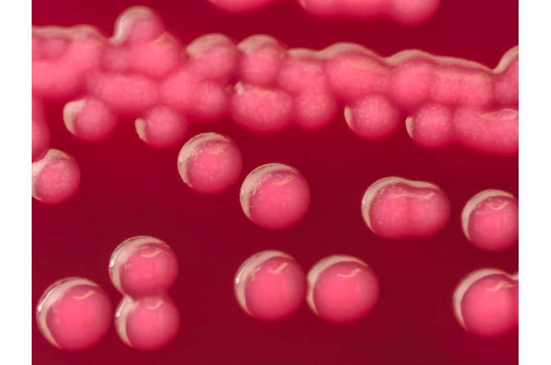
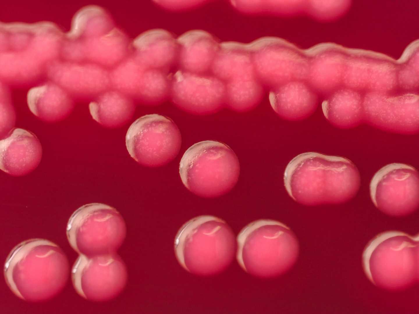
In this study, researchers from UCL developed three-dimensional cell models capable of mimicking the biological environment and function of human bladder tissue, in order to observe the interactions between host and pathogen in conditions as close to the human body as possible. These ‘mini bladders’ were exposed to six bacterial species commonly found in the human bladder: Escherichia coli, Enterococcus faecalis, Pseudomonas aeruginosa, Proteus mirabilis, Streptococcus agalactiae and Klebsiella pneumoniae.
Professor Jennifer Rohn, senior author of the study from UCL Division of Medicine, said, “We put a variety of UTI bacteria species and strains through their paces and discovered a battleground of diversity. One of the key observations was the importance of persistence. If you want to be a successful pathogen, you have to have strategies that help you to survive treatment and hide from patrolling immune cells, which means you live to fight another day.”
“Some species of both ‘good’ and ‘bad’ bugs formed pods within the bladder wall, most likely as a way of surviving in this harsh environment. If this happens with a friendly bug, this isn’t a problem. But if the bug is causing an infection, this poses a serious problem for diagnosis and treatment because the bacteria aren’t necessarily going to be detected in a urine sample or be in a position where oral antibiotics can reach them.”
The study also found that human cells were very good at distinguishing friendly from not-so-friendly bacteria, regardless of whether they could invade the bladder wall or not. All the ‘bad’ bugs tested triggered the production of immune molecules, called cytokines, and the shedding of the top layer of the bladder wall, whereas the ‘good’ bacteria could colonize the bladder wall without triggering an immune response.
Dr. Carlos Flores, first author of the study from UCL Division of Medicine, said, “Based on our results, next-generation diagnostics for UTIs could focus on identifying ‘bad’ bugs based on how the body responds, rather than trying to spot the presence of problem bacteria among the background noise of the microbiome. There are so many species and strains of bacteria in the human bladder that we don’t fully understand, but the body seems to be pretty good at telling friend from foe.”
The findings indicate that effective treatments for persistent UTIs may require the ability to penetrate human tissues, in order to reach bacterial populations dwelling in the bladder wall. A UCL spin-out, AtoCap, is currently developing ways to deliver drugs inside cells to target pathogens hiding there.
Professor Rohn concluded, “This study confirms what many women who’ve struggled with persistent UTIs already know, which is that the current methods of diagnosing and treating these infections are inadequate.”
“Urine dipstick tests are too likely to miss infections hiding in the bladder wall, especially when a patient’s first response to discomfort is to drinks lots of water, which dilutes the test. Not all bugs can be cultured in the lab, and even if they could be that doesn’t tell us if this strain is the cause of an infection or if its position in the bladder wall would make the standard three-day course of antibiotics unlikely to eradicate it.”
Carolyn Andrew, Director of the Chronic Urinary Tract Infection Campaign (CUTIC), said, “This research has been instrumental in providing unequivocal evidence for our national campaign to improve testing and diagnosis of chronic, persistent UTIs. Professor Rohn’s work in this field is a vitally important step forwards and should help tens of thousands of women in the UK to receive effective diagnosis and treatment of a chronic infection in their bladders.”
The mini bladders used in the study are currently the most sophisticated artificial bladder models in the world. About the size of a five-pence coin (UK) or a dime (US), the models are grown in plastic dishes bathed in human urine.
Unlike other models, the mini bladders have seven to eight layers to resemble the structure of the human bladder and can operate in 100% urine, a toxic environment that is the natural habitat of UTI bugs. They include lifelike human features such as a mucous-like barrier that separates the bladder wall from urine and the ability to emit immune distress signals when attacked.
More information:
Carlos Flores et al, A human urothelial microtissue model reveals shared colonization and survival strategies between uropathogens and commensals, Science Advances (2023). DOI: 10.1126/sciadv.adi9834. www.science.org/doi/10.1126/sciadv.adi9834
Journal information:
Science Advances
Source: Read Full Article
