In a recent article posted to the bioRxiv* preprint server, investigators analyzed whether Mexican free-tailed bats were a severe acute respiratory syndrome coronavirus 2 (SARS-CoV-2) reservoir.

Background
SARS-CoV-2, which arose from Asian wild bats, has caused the Coronavirus disease 2019 (COVID-19) pandemic. Numerous concerns about the ecology of the virus remain unresolved as the pandemic enters its third year.
It is crucial to understand how SARS-CoV-2 interacts with wildlife, such as whether 1) North American wildlife can serve as viral reservoirs; 2) the SARS-CoV-2 can adapt genetically to novel hosts and become more virulent; or 3) the virus can impact the health of wild populations, especially endangered or threatened species.
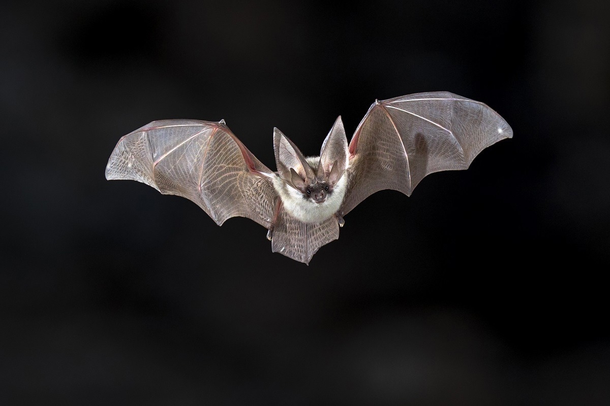
Notably, concerns have been expressed that SARS-CoV-2 would spread to new hosts and infect bats and other wildlife species in North America. Large colonies of Mexican free-tailed bats (Tadarida brasiliensis (TABR)) are found in the southern United States (US), frequently in populated regions. As a result, they may come into contact with SARS-CoV-2 through infected people. Since they are migratory species, this species may carry SARS-CoV-2 from or to South and Central America if they are susceptible to it.
About the study
In the present study, the researchers experimentally infected wild TABR with SARS-CoV-2 to assess the reservoir possibility, susceptibility, population effects of infection among this species, and adaptability of the virus to a new probable host.
The team conducted reverse transcription-polymerase chain reaction (RT-PCR) assessments of fecal material assembled from the TABR before the viral challenge. They used quantitative RT-PCR (qRT-PCR) examination of oral and rectal swabs to evaluate the SARS-CoV-2 excretion following experimental virus inoculation of the bats.
A competitive enzyme-linked immunosorbent assay (cELISA) was utilized to detect SARS-CoV-2 antibodies. The scientists monitored clinical signs of SARS-CoV-2 infection throughout the three-week study duration among the animals. They also conducted histopathologic and postmortem examinations.
Besides, immunohistochemistry was used to find the SARS-CoV-2 antigen in sections from the rostral lung, nasal cavity, heart, liver, spleen, pancreas, small and large intestine, stomach, and brain from a total of 14 mock and experimentally inoculated bats. In addition, the authors conducted qRT-PCR analyses of tissues gathered from the control and SARS-CoV-2-infected bats.
Results and discussions
Overall, the study results indicated that five of the ten oronasally SARS-CoV-2-inoculated bats developed the infection and orally emitted a moderate amount of virus for six to 18 days following the viral challenge. Before the study's completion, all five animals mounted an immune reaction, seroconverted, and cleared the virus with no overt signs of illness. Additionally, the team discovered no indications of virus transmission to uninoculated cohoused TABR.
The utilized inoculum titer, i.e., 105 median tissue culture infectious dose (TCID50)/dose, was probably close to the 50% infectious dose for TABR since five of the ten, or 50%, inoculated bats were virus-infected. Yet, the highest viral concentration excreted by infected bats was within 103 to 104 TCID50 equivalents/ml. Therefore, contact transmission of SARS-CoV-2 between TABR would be improbable based on these data.
The way the researchers kept the bats during the challenge phase was another potential explanation for why there was no viral transfer between TABR. In the wild, TABR roosts in large colonies in anthropogenic and natural structures such as caves, bridges, and culverts. One infected bat and one uninoculated bat were cohoused in cages of around 1 meter3 for this investigation. The bats were not forced to cluster as densely in this relatively vast area as they would have in a natural roost, perhaps preventing the spread of the virus.
The study also discovered an absence of viral elimination through the digestive system, apart from the evident lack of transmission. The authors found no proof of virus in any bats' digestive tracts or rectal swabs, including the infected bats.
Another observation was that the SARS-CoV-2 infection in TABR did not appear to have any noticeable adverse health impacts. Although it was uncertain if infected wild bats would have impaired ability to provide maternal care, scavenge for food, or conduct other life activities, the research findings suggest that TABR populations were probably not at risk from the COVID-19 pandemic.
Conclusions
The present findings showed that although TABR was vulnerable to SARS-CoV-2 infection, it might not lead to mortality among infected wild TABR populations. Nevertheless, SARS-CoV-2 transmission from TABR to or from humans and other animal species is a definite possibility that needs more research for proper definition. The team noted that a crucial follow-up investigation would involve accurately estimating the SARS-CoV-2 infectious dosage in TABR.
The current study data was beneficial for wildlife biologists, bat rehabilitationists, cave recreationists, and the common public if they engage with Mexican free-tailed bats or go into caves or other roosting environments of the bats.
*Important notice
bioRxiv publishes preliminary scientific reports that are not peer-reviewed and, therefore, should not be regarded as conclusive, guide clinical practice/health-related behavior, or treated as established information.
- Jeffrey Hall, Erik Hofmeister, Hon Ip, Sean Nashold, Ariel Leon, Carly Malave, Elizabeth Falendysz, Tonie Rocke, Mariano Carossino, Udeni Balasuriya, Susan Knowles. (2022). Experimental infection of Mexican free-tailed bats (Tadarida brasiliensis) with SARS-CoV-2. bioRxiv. doi: https://doi.org/10.1101/2022.07.18.500430 https://www.biorxiv.org/content/10.1101/2022.07.18.500430v2
Posted in: Medical Science News | Medical Research News | Disease/Infection News
Tags: Antibodies, Antigen, Assay, Brain, Coronavirus, Coronavirus Disease COVID-19, covid-19, Digestive System, Enzyme, Food, Heart, Immunohistochemistry, Large Intestine, Liver, Mortality, Pancreas, Pandemic, Polymerase, Polymerase Chain Reaction, Research, Respiratory, SARS, SARS-CoV-2, Severe Acute Respiratory, Severe Acute Respiratory Syndrome, Spleen, Stomach, Syndrome, Tissue Culture, Transcription, Virus

Written by
Shanet Susan Alex
Shanet Susan Alex, a medical writer, based in Kerala, India, is a Doctor of Pharmacy graduate from Kerala University of Health Sciences. Her academic background is in clinical pharmacy and research, and she is passionate about medical writing. Shanet has published papers in the International Journal of Medical Science and Current Research (IJMSCR), the International Journal of Pharmacy (IJP), and the International Journal of Medical Science and Applied Research (IJMSAR). Apart from work, she enjoys listening to music and watching movies.
Source: Read Full Article